Naoya Shibata shares his research at Science Lecture
2023-11-02
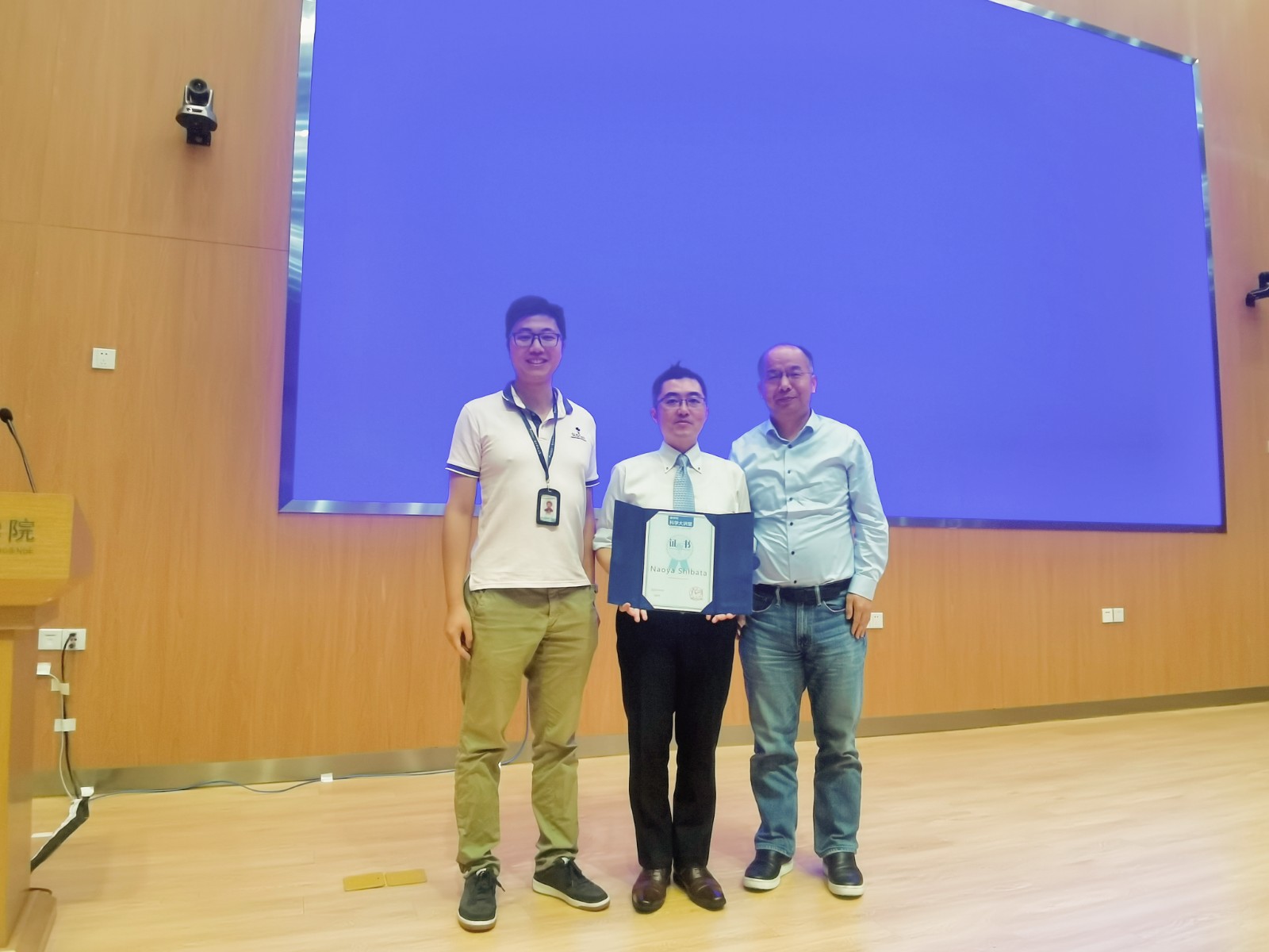
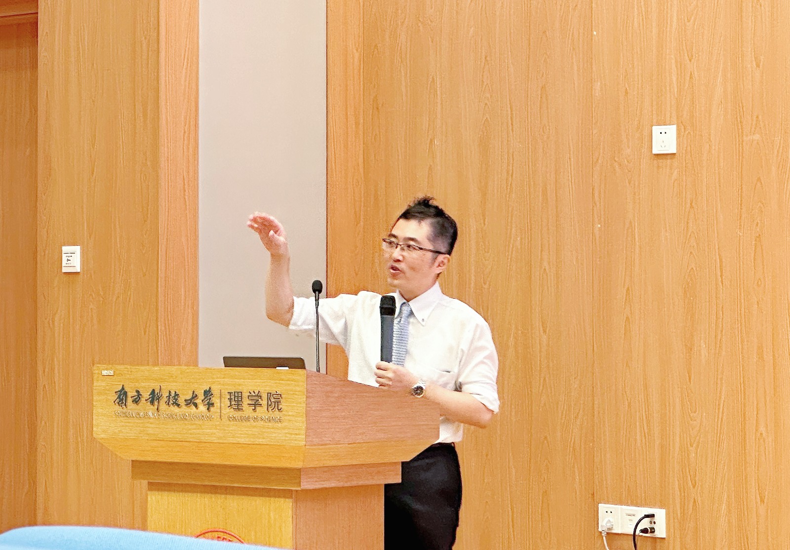
On October 30, 2023, Prof. Naoya Shibata from the University of Tokyo was invited to the 133 Science Lecture in the College of Science. He gave a lecture themed “Magnetic-field-free Atomic Resolution STEM”. Jiaqing HE, Associate Dean of the College of Science, and Junhao LIN, Associate Professor of the Department of Physics at SUSTech handed an honorary certificate to Prof. Naoya Shibata.

Prof. Shibata introduced the functional advantages of transmission electron microscopy, such as sub-angstrom resolution, ABF imaging of light atoms, and strong EELS/EDS element characterization. He then pointed out that it is very difficult to balance low electron dose and high signal-to-noise ratio for electron beam-sensitive samples. To address this challenge, Prof. Shibata's team recently developed an optimized bright field (OBF) scanning transmission electron microscopy technique for low electron dose imaging. It can reconstruct images with a high signal-to-noise ratio and dose efficiency about two orders of magnitude higher than traditional methods. It acts on typical electron irradiation-sensitive zeolite materials, effectively visualizing all atomic sites in the porous structure framework.

Subsequently, Prof. Shibata introduced the design concept and case application of using differential phase contrast scanning transmission electron microscopy (DPC-STEM) to directly observe the electric and magnetic fields at the interface between materials and devices. Due to changes in local scattering conditions throughout the entire sample range, the intensity fluctuates in the bright field image mode. This fluctuation in local scattering intensity is superimposed with the effect of the electric field, thereby weakening the observation effect of the electric field. Based on DPC-STEM technology, tDPC-STEM technology has been developed. This technology averages the results of multiple beam tests and extracts the true electromagnetic field signal using the differential phase contrast signal obtained from the experiment. It can suppress diffraction contrast and improve the imaging effect of two-dimensional electron gas in GaN HEMT heterogeneous interfaces. In order to achieve visualization of the spatial distribution of magnetic fields inside magnetic materials, Prof. Shibata's team utilized a front and rear anti-symmetric lens design with opposite polarity. The magnetic fields on the sample plane can cancel each other out and decrease to zero, enabling the sample to achieve original sub-level resolution characterization in a magnetic field-free environment. Taking α-Fe2O3 as an example, although the atomic scale magnetic field signal is extremely weak, the real space visualization of α-Fe2O3 intrinsic magnetic field is achieved by accurately subtracting the atomic electric field and averaging the single cell image to improve the signal-to-noise ratio.
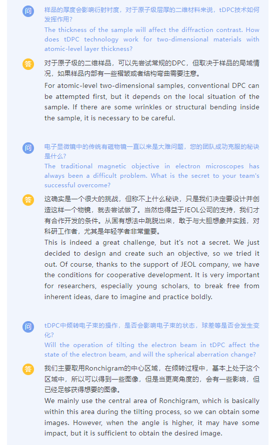
During the Q&A session, the on-site teachers and students enthusiastically asked questions about tDPC technology, magnetic field-free electron microscopy, and other aspects. Prof. Shibata provided detailed answers, and the on-site discussion was lively.


